
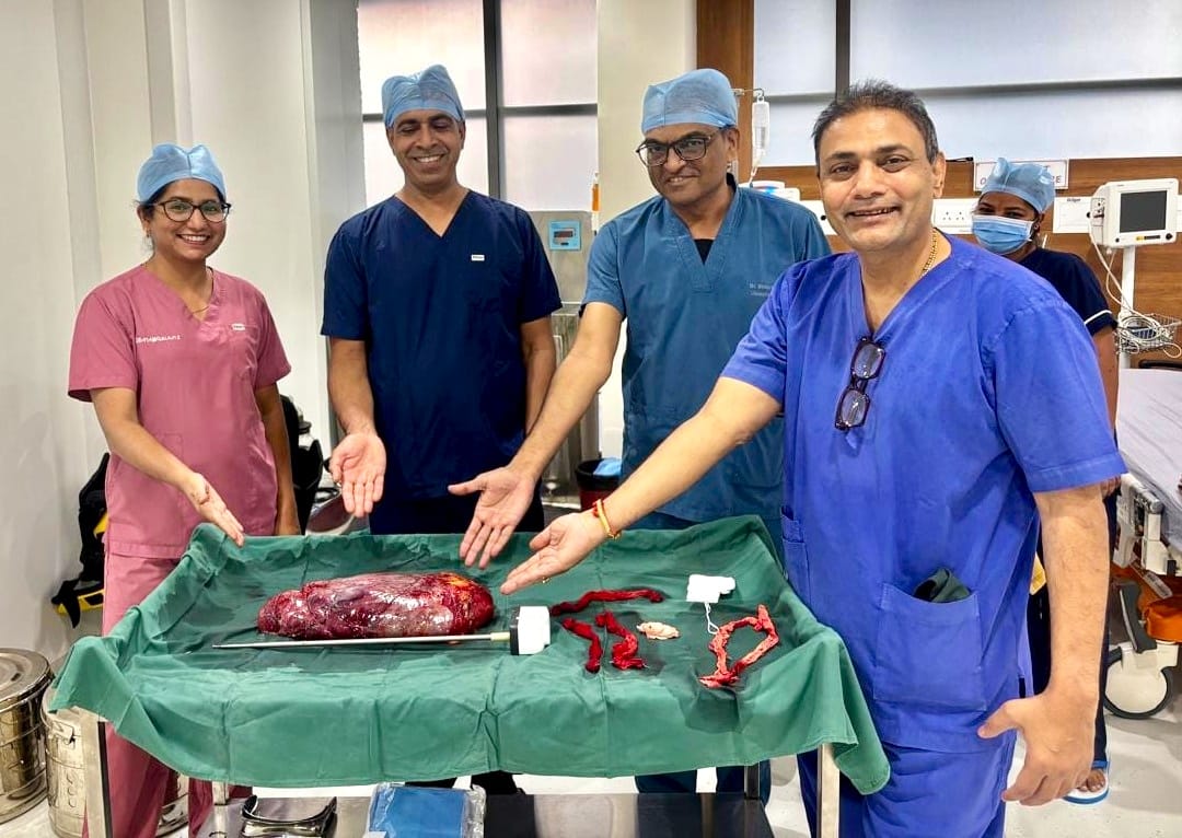
Ahmedabad: In a rare and technically demanding surgical achievement, Dr. Dipak Limbachiya and the surgical team at Eva Women’s Hospital have successfully removed a massive 30-centimetre retroperitoneal tumour from a 41-year-old woman using advanced laparoscopic (minimally invasive) surgery.

Despite the tumour partially encasing and displacing vital organs and major blood vessels—including the kidney, liver, aorta, and inferior vena cava (IVC)—the surgical team achieved complete removal of the mass with zero intraoperative complications and no damage to surrounding organs.
Patient Background and Clinical Presentation
The patient, a 41-year-old woman, sought medical evaluation after experiencing progressive abdominal swelling and distension over an eight-month period.
Advanced radiological imaging identified a massive growth measuring approximately ~30 x 19 x 12 cm located deep in the retroperitoneum—the space situated behind the abdominal lining. An ultrasound-guided core needle biopsy diagnosed the lesion as an atypical lipomatous tumour.
The mass had reached a critical size and position:
Organ Displacement: It partially encased and displaced the right kidney and the main renal blood vessels.
Critical Anatomical Span: It extended from deep within the pelvis upward to the right hemidiaphragm and the back surface of the right liver lobe.
Vascular Proximity: It reached centrally into the pre-aortic, precaval, and left para-aortic spaces, sitting directly against the body’s primary blood pathways (the aorta and vena cava).
Surgical Innovation: Why This Case Is Exceptional
Tumours of this magnitude in the retroperitoneum are almost universally treated through large, open abdominal incisions because of the severe risk of catastrophic bleeding or organ injury.
Operating in this tight, deep anatomical corridor using minimally invasive keyhole instruments required exceptional precision and advanced tissue separation (laparoscopic-assisted adhesiolysis). Dr. Limbachiya meticulously dissected the tumour off the major blood vessels and preserved the function of all adjacent organs, offering the patient the benefits of minimally invasive surgery: less trauma, reduced pain, and a faster pathway to recovery.
“Operating within the retroperitoneal space is among the most challenging frontiers in surgery because it houses life-critical vascular highways,” said Dr. Dipak Limbachiya. “When a tumour reaches 30 centimetres and wraps closely around the kidney, liver, and aorta, open surgery is usually the default. Achieving complete clearance laparoscopically without a single vascular injury demonstrates how precision keyhole surgery can transform outcomes even in high-risk abdominal oncology.”
Medical Facts & Rarity at a Glance
Low Global Incidence: Retroperitoneal tumours are exceptionally rare, diagnosed in only 0.3 to 0.4 people per 100,000 annually (approximately 2.7 cases per million individuals).
Rare Cancer Category: Primary retroperitoneal sarcomas account for only 10% to 15% of soft-tissue tumours and represent less than 1% of all adult malignancies.
Surgical Rarity: While standard laparoscopic removal of small fatty tumours is documented, worldwide medical literature reports very few cases successfully managed laparoscopically at this level of size and vascular complexity.
Media Backgrounder: What Is a Retroperitoneal Mass?
Definition: An abnormal solid or semi-solid growth located behind the peritoneum, the membrane lining the abdominal wall. This compartment contains vital structures like the kidneys, aorta, and inferior vena cava.
“Silent” Symptoms: Because the abdominal cavity can expand, these tumours often produce no symptoms until they become massive. Common warning signs include progressive abdominal enlargement, vague abdominal or lower back pain, early fullness during meals, digestive disruption, or unexplained swelling in the legs.
Diagnostic Protocol: Contrast-enhanced CT scans and MRI imaging are essential to determine tumour boundaries, followed by image-guided needle biopsy for definitive tissue classification.